

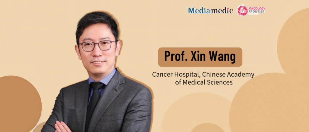
The Samsung Club National Young Breast Cancer Experts Conference recently concluded in Zhengzhou, bringing together rising leaders from China's four major breast cancer academic organizations to exchange ideas on precision oncology, physician training, and multidisciplinary collaboration.Following the meeting, Oncology Frontier interviewed Professor Xin Wang of the National Cancer Center/National Clinical Research Center for Cancer/Cancer Hospital, Chinese Academy of Medical Sciences (CAMS). In this conversation, he discusses the evolution of breast cancer surgery, the increasingly central role of imaging in precision surgical decision-making, and why mastering imaging should become an essential competency for the next generation of breast surgeons.
Oncology Frontier: Could you give us an overview of the current state of breast cancer surgery and where the field is headed?
Professor Xin Wang:
Looking back over the history of breast surgery, the overall direction has been one of continuous de-escalation. Surgical treatment has evolved from radical mastectomy and extended radical mastectomy to modified radical mastectomy and, ultimately, to breast-conserving surgery. Throughout this evolution, the extent of tissue resection has steadily decreased.
Management of the axilla has followed a similar trajectory. Routine axillary lymph node dissection gave way to sentinel lymph node biopsy, and more recently, appropriately selected patients can even avoid sentinel lymph node biopsy altogether.
There have, of course, been important developments that move in a different direction, such as breast reconstruction. Although breast surgery has generally become less extensive, reconstruction often involves more complex procedures. For patients who are not candidates for breast-conserving surgery, reconstruction using autologous tissue or implants has become an increasingly important component of treatment. At the same time, minimally invasive techniques—including endoscopic and robotic surgery—have expanded the surgical options available to both physicians and patients.
Overall, the future of breast surgery is clear: operations are becoming increasingly conservative and increasingly precise. Tissue that requires removal should be completely excised, while healthy tissue that does not need to be removed should be preserved whenever possible. As for how that goal is achieved, the field is currently experiencing remarkable innovation, with multiple surgical techniques continuing to evolve.
Oncology Frontier: From mammography and ultrasound to advanced multiparametric MRI, imaging has become increasingly sophisticated. In the era of precision surgery, what is the greatest value of imaging in helping surgeons decide how much to remove, how to operate, and when surgery should be performed?
Professor Xin Wang:
This is an extremely important question, yet one that is often overlooked.
Cancer management always begins with diagnosis before treatment. Patients first undergo a physical examination, followed by various diagnostic tests, among which imaging is arguably the most important. Ultrasound, mammography, MRI, and PET-CT all play indispensable roles in modern breast cancer care, although each modality has its own strengths and limitations. Mammography performs less well in women with dense breasts, ultrasound can be more challenging in patients with larger breasts, and MRI is associated with a relatively high false-positive rate.
For this reason, breast surgeons themselves must develop a solid understanding of imaging. During residency training, I believe every surgical trainee should complete a rotation in radiology. I also encourage young surgeons to review imaging studies independently. They are not expected to reach the same level of expertise as radiologists, but they should understand how different types of breast tumors appear on ultrasound, mammography, and MRI, and they should recognize conditions that can mimic malignancy. These are fundamental clinical skills for every breast surgeon.
Today, artificial intelligence is rapidly transforming imaging analysis. Image interpretation is gradually shifting from what might be called “human estimation” toward computer-assisted prediction. Will artificial intelligence ultimately be more accurate? That depends on the quality of the data used to train it.
Our own team has been actively involved in this area. Before machine-learning algorithms can make accurate predictions, radiologists and surgeons must first annotate tumor location, size, and extent. Once those data are available, AI can analyze images more rapidly and, in many cases, more accurately.
These are precisely the parameters that determine surgical planning. Surgeons must know where the tumor is located, how large it is, whether it is confined to one focus or involves multiple lesions, and what its true boundaries are. These factors directly determine how surgery should be performed.
Combined with artificial intelligence, imaging will increasingly support surgical navigation, facilitate more accurate definition of tumor margins, optimize the extent of resection, and even assist with reconstructive planning through technologies such as three-dimensional printing.
Imaging is equally important when determining when surgery should be performed. Following neoadjuvant therapy, imaging allows us to evaluate the degree of tumor shrinkage, identify residual disease, distinguish between concentric and scattered patterns of tumor regression, and determine whether the planned resection can safely be reduced. These are all critical pieces of information that directly influence surgical decision-making.
Every imaging modality has advantages and limitations. With the addition of artificial intelligence, however, imaging is becoming an even more powerful tool for precision surgery. For young breast surgeons, learning to interpret imaging is no longer optional—it is an essential professional skill.
Oncology Frontier: Young surgeons often devote most of their training to operative techniques, while spending relatively little time developing expertise in imaging. What advice would you give young breast surgeons who want to strengthen these interdisciplinary skills during busy clinical training?
Professor Xin Wang:
As I mentioned earlier, imaging interpretation is an essential competency for breast surgeons.
Traditionally, surgeons have been fascinated by the technical aspects of surgery itself. Every surgeon remembers the excitement of placing their first suture, making their first skin incision, or removing their first tumor. Those milestones are important, but they should come only after mastering diagnosis.
Without an accurate diagnosis, there can be no accurate treatment.
That is why I strongly believe that every surgical trainee should complete a dedicated rotation in radiology and become familiar with the imaging characteristics of different breast lesions.
Consider a simple example. If a patient brings imaging studies to your clinic and you are unable to identify the tumor’s size or extent, it will inevitably undermine the patient’s confidence in your recommendations.
Physical examination and imaging interpretation form the foundation of surgical decision-making. Once these fundamental skills are firmly established, learning operative techniques becomes a natural progression. There is no need to rush into surgery before developing the diagnostic skills that guide it.
For young surgeons, building these fundamentals will ultimately create far greater opportunities for professional growth throughout their careers.
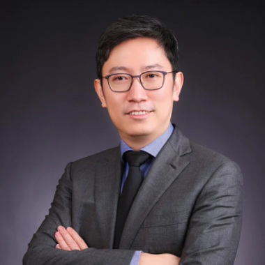
Professor Xin Wang
National Cancer Center / National Clinical Research Center for Cancer / Cancer Hospital, Chinese Academy of Medical Sciences (CAMS)