
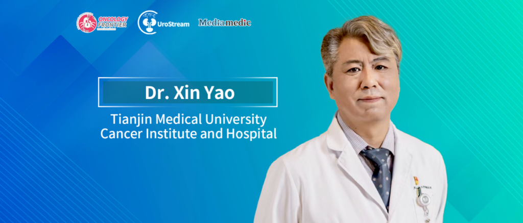
Editor's Note: With the rapid development of molecular imaging, artificial intelligence, multi-omics technologies, and robotic surgery, the management of urologic cancers is gradually evolving from relatively standardized treatment toward more precise, minimally invasive, and individualized whole-course care. Precision medicine can improve the accuracy of tumor identification, risk stratification, and treatment selection, while robotic-assisted surgery provides important technical support for meticulous dissection, organ preservation, and functional reconstruction. Oncology Frontier · UroStream invited Professor Xin Yao of Tianjin Medical University Cancer Institute and Hospital for an exclusive interview to discuss pathways toward precision diagnosis and treatment in urologic oncology, as well as the clinical value and future directions of robotic surgery.Integrating Imaging, Pathology, and Multi-Omics Data to Drive More Precise Clinical Decision-Making
Oncology Frontier · UroStream: As precision medicine continues to evolve, the management of urologic cancers is becoming increasingly individualized. In your view, how can we further advance precision diagnosis and treatment in urologic oncology?
Professor Xin Yao: Precision diagnosis and treatment has become a major focus across oncology in recent years. It encompasses two key dimensions: precision diagnosis and precision treatment. Its fundamental goal is to understand each patient’s disease characteristics as accurately as possible and, based on that understanding, select the most appropriate treatment strategy.
In terms of precision diagnosis, imaging technologies are advancing rapidly. Clinical diagnosis has expanded from conventional anatomical imaging such as CT and MRI to PET/CT and molecular imaging targeting specific molecular markers. Taking prostate cancer as an example, the application of PSMA PET/CT has improved the detection of primary lesions, lymph-node metastases, and distant metastases, providing more comprehensive information for disease staging, localization of recurrence, and treatment planning.
In the future, artificial intelligence will further enhance the use of imaging and pathological information. AI-assisted imaging interpretation and pathological diagnosis have already demonstrated considerable potential. By analyzing high-dimensional features in medical images and pathological slides, radiomics and pathomics may extract information that is difficult to identify through conventional visual assessment and apply it to tumor classification, risk prediction, and treatment response assessment.
However, the value of artificial intelligence lies not simply in accelerating diagnosis. More importantly, it can help clinicians integrate complex information. If imaging characteristics, pathological morphology, genomics, transcriptomics, and clinical parameters can be effectively integrated in the future, we may be able to construct a more comprehensive disease profile and move from simply determining “whether a tumor is present” toward understanding “what biological behavior the tumor has and which treatments it is likely to respond to.”
In terms of precision treatment, treatment options for urologic cancers are becoming increasingly diverse, including surgery, radiotherapy, targeted therapy, immunotherapy, endocrine therapy, and various combination strategies. The growing number of treatment options provides patients with more opportunities, but it also makes clinical decision-making more complex.
We need to determine not only whether a particular treatment is effective, but also which patients are most likely to derive a meaningful survival benefit, who may be at greater risk of severe adverse events, and how different treatments should be sequenced and combined. A single clinical parameter is often insufficient to answer these complex questions.
Therefore, an important future pathway for precision treatment is to integrate high-quality clinical data and large-scale real-world data, incorporating multiple dimensions such as age, performance status, disease stage, pathological characteristics, genomic alterations, imaging findings, previous treatments, and treatment-related adverse events. These data can then be used to develop clinically interpretable predictive models.
Such models must undergo rigorous validation and ultimately address concrete clinical questions—for example, whether a patient requires treatment intensification, whether monotherapy or combination therapy is more appropriate, when treatment should be modified, and how to balance efficacy and safety.
True precision treatment is not simply about adding more tests or treatment options. It is about choosing the right treatment for the right patient at the right time.
Harnessing the Advantages of Robotic Platforms to Move Cancer Surgery from Minimally Invasive Treatment toward Functional Preservation
Oncology Frontier · UroStream: In recent years, robotic-assisted surgery and minimally invasive technologies have developed rapidly in urology. What changes have these technologies brought to clinical practice, and what breakthroughs are still needed?
Professor Xin Yao: Over the past decade or more, driven by leading Chinese urologists including Academician Xu Zhang, robotic and minimally invasive surgery in China has developed rapidly. Significant progress has been made in complex urologic procedures, remote surgery, and the development of standardized technical systems. In some areas, we have even reached an internationally leading position.
In certain highly complex procedures, such as minimally invasive treatment of renal cell carcinoma with inferior vena cava tumor thrombus, Chinese teams have accumulated extensive experience. Exploration of remote robotic surgery has also created new possibilities for overcoming geographical barriers to medical resources and extending access to high-quality surgical expertise.
At the same time, the concept of functional preservation has received increasing attention. This has encouraged the surgical management of prostate cancer and other urologic malignancies to evolve from focusing solely on tumor removal toward balancing oncologic control with functional preservation.
One of the major advantages of robotic surgical systems is their ability to provide a magnified, high-definition three-dimensional surgical view and facilitate meticulous dissection, cutting, hemostasis, and reconstruction through flexible and stable instruments.
Compared with open surgery, robotic procedures are generally less invasive. Compared with conventional laparoscopy, robotic systems offer greater flexibility and suturing capability, particularly in confined anatomical spaces. These characteristics are especially well suited to urologic surgery.
Whether performing tumor excision and renal reconstruction during partial nephrectomy or preserving the neurovascular bundles, reconstructing the urethra, and managing the bladder neck during radical prostatectomy, surgeons need to identify and manipulate delicate anatomical structures with great precision.
As robotic technology becomes more widely adopted, an increasing number of appropriately selected patients with renal tumors can undergo nephron-sparing surgery, allowing clinicians to control the tumor while preserving as much normal renal function as possible.
For patients with prostate cancer, surgical treatment is also placing greater emphasis on postoperative urinary continence and sexual function. Through meticulous dissection and functional reconstruction, some patients can achieve better perioperative recovery and long-term quality of life.
Therefore, the impact of robotic surgery goes beyond changes in incision size or surgical technique. More importantly, it is helping to transform the goals of surgical treatment. Modern cancer surgery should not focus solely on whether the operation can be completed successfully; it must also consider oncologic safety, organ function, postoperative recovery, and long-term quality of life.
Looking ahead, robotic surgery will continue to focus on improving precision and functional preservation.
On the one hand, surgical techniques and standardized procedures should continue to be optimized so that complex operations can be performed more safely and consistently. On the other hand, robotic platforms need to be integrated more closely with intraoperative imaging, fluorescence navigation, molecular imaging, and artificial intelligence.
At present, surgeons still rely heavily on what they can see with their eyes and on their individual experience. In the future, if preoperative imaging can be fused with the intraoperative surgical field in real time, while AI can help identify tumor boundaries, vascular anatomy, and critical functional structures, we may be able to overcome some of the limitations of conventional visualization and experience.
This could enable surgery to progress from “precision in execution” toward “precision in recognition” and ultimately “precision in decision-making.”
At the same time, the development of robotic technology should not focus solely on increasingly sophisticated equipment. It must be accompanied by improvements in surgeon training, quality control, and standardized clinical application. Only by establishing comprehensive training systems, surgical evaluation frameworks, and appropriate patient-selection criteria can new technologies be translated into stable and reproducible clinical benefits.
Overall, precision medicine and robotic minimally invasive technology are not two independent pathways. Precision diagnosis helps clinicians identify disease characteristics, assess risk, and select appropriate treatments, while robotic technology enables surgeons to execute those treatment strategies with greater precision.
Their coordinated development has the potential to move urologic oncology further from experience-driven care toward data-driven decision-making and precision treatment, while maintaining tumor control and maximizing preservation of patients’ organ function and quality of life.

Professor Xin Yao