

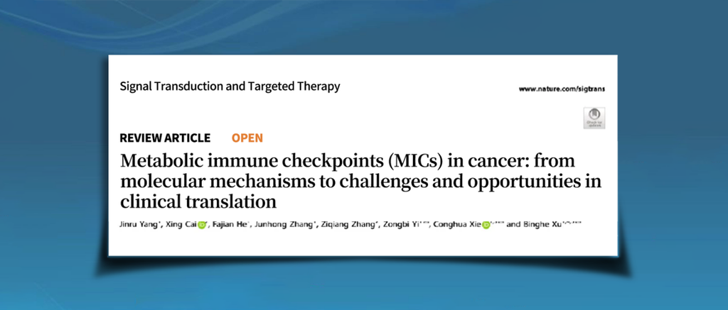
On October 9, 2026, a research team from Zhongnan Hospital of Wuhan University, the Taikang Center for Life and Medical Sciences at Wuhan University, the National Cancer Center, and the Cancer Hospital of the Chinese Academy of Medical Sciences published a review article online in Signal Transduction and Targeted Therapy (impact factor: 81.2), entitled “Metabolic immune checkpoints (MICs) in cancer: from molecular mechanisms to challenges and opportunities in clinical translation.” The review was co-corresponding-authored by Academician Qianjun Xu, Professor Xiehua Xie, and Professor Yechun Yi. It systematically examines the molecular mechanisms through which tumor metabolic reprogramming drives immune evasion and proposes a theoretical framework centered on metabolic immune checkpoints (MICs), offering a new conceptual basis for understanding immunotherapy resistance and developing combined metabolic and immunotherapeutic strategies.Immune checkpoint inhibitors have profoundly transformed cancer treatment, yet many patients fail to achieve durable benefits, and some initially responsive patients develop resistance early in treatment. Traditionally, immunotherapy failure has been explained primarily through classical immune checkpoints such as PD-1/PD-L1 and CTLA-4, as well as T-cell exhaustion and deficiencies in tumor antigens. However, emerging evidence indicates that nutrient competition, metabolite accumulation, and hypoxia-driven metabolic changes in the tumor microenvironment can substantially affect the survival, differentiation, and effector functions of immune cells. Through metabolic reprogramming, tumor cells not only meet their own proliferative demands but also establish a metabolic ecosystem in their surroundings that favors immune evasion.
The Tumor Metabolism–Immune Regulation Network
The authors first establish a tumor metabolism–immune regulation network and classify MICs into three categories according to their mechanisms of action.
MIC-I primarily refers to metabolic enzymes or metabolites that, after post-translational modifications such as lactylation, glycosylation, and acetylation, regulate the expression and function of classical immune checkpoints, including PD-1/PD-L1.
MIC-II refers to metabolic regulatory checkpoints involving metabolic enzymes, metabolites, and nutrient competition that directly influence immune-cell function.
MIC-III emphasizes the systemic effects of the gut microbiota and its metabolites on antitumor immunity.
This framework extends the traditional concept of immune checkpoints beyond receptor–ligand interactions on cell surfaces to encompass metabolic enzymes, nutrients, metabolites, microorganisms, and their involvement in regulating the tissue microenvironment. It recognizes that tumor immune evasion does not occur solely through interactions between immune cells and tumor cells; it also develops through competition for nutrients, alterations in the metabolic environment, and the remodeling of local ecosystems. MICs therefore represent important hubs linking tumor metabolism, immune function, and treatment response.
MIC-I: Metabolic Modifications Regulate Classical Immune Checkpoints
For MIC-I, the central mechanism is that the metabolic state of tumor cells can alter the expression and function of classical immune checkpoints through multiple post-translational modifications.
Enhanced glycolysis, lipid synthesis, amino acid metabolism, and hexosamine biosynthesis in tumor cells can generate metabolic intermediates, including lactate, acetyl-CoA, acetylated metabolites, and UDP-GlcNAc. These molecules not only participate in energy production and biosynthesis but can also provide substrates for post-translational modifications of proteins, thereby affecting the stability, cellular localization, and functional activity of immune checkpoint molecules such as PD-L1.
For example, glycosylation, lactylation, phosphorylation, acetylation, palmitoylation, and ubiquitination can influence the regulation of PD-L1. These modifications may affect…
These modifications can affect PD-L1 stability on the cell membrane, its degradation, and its interactions with other signaling proteins. At the same time, signaling pathways such as NF-κB, JAK/STAT3, and AMPK are also regulated by metabolic states, further promoting the expression of immunosuppressive molecules. Through these mechanisms, tumor cells can convert metabolic alterations into persistent immunosuppressive signals, impairing the antitumor functions of T cells, NK cells, and other immune cells. This suggests that future immunotherapy may need to target not only classical immune checkpoints but also the metabolic regulatory processes underlying their activity.
MIC-II: Nutrient Competition and Metabolic Stress Impair Immune Cell Function
Whereas MIC-I primarily focuses on how metabolic states modify and regulate immune checkpoint molecules, MIC-II further reveals how tumor cells directly weaken immune-cell function through nutrient competition and metabolic stress.
Tumor cells typically have a greater capacity to take up glucose, glutamine, and fatty acids, competing with T cells, NK cells, and dendritic cells for limited nutrients in the tumor microenvironment. When immune cells cannot obtain sufficient glucose, amino acids, or lipids, their proliferation, cytotoxicity, and cytokine production are impaired.
Meanwhile, metabolites released by tumor cells, including lactate, reactive oxygen species, ammonia, and urea-cycle metabolites, can create an acidic, hypoxic, and oxidatively stressful microenvironment. This environment further disrupts mitochondrial function and energy metabolism in immune cells. As a result, some T cells gradually enter a state of functional exhaustion, NK-cell killing capacity declines, and antigen-presenting cells are also impaired.
In contrast, regulatory T cells, tumor-associated macrophages, and myeloid-derived suppressor cells can often adapt more effectively to this metabolic environment, further reinforcing immunosuppression. This “metabolic immune barrier,” formed by nutrient availability, metabolic byproducts, mitochondrial function, and intercellular metabolic interactions, therefore constitutes an important foundation for tumor immune evasion.
MIC-III: Gut Microbial Metabolites Participate in the Regulation of Antitumor Immunity
Beyond tumor cells and the local microenvironment, the review extends its scope to the gut microbial ecosystem and proposes MIC-III, a regulatory model involving microbial metabolites and immune responses.
Gut microorganisms produce short-chain fatty acids, bile acids, tryptophan-derived metabolites, polyamines, and other biologically active compounds. These substances can influence the tumor microenvironment through the circulation or relevant immune signaling pathways, regulating immune-cell differentiation, migration, metabolic adaptation, and effector functions.
For example, short-chain fatty acids can regulate the functions of regulatory T cells and bone marrow-derived cells through specific receptors. Tryptophan-derived metabolites can influence regulatory and effector T cells through the aryl hydrocarbon receptor (AhR). Bile acids and polyamine metabolites of microbial origin may alter NK-cell activity, CD8+ T-cell cytotoxicity, and local inflammatory states.
The review also summarizes the potential roles of Bifidobacterium, Lactobacillus, and other bacterial communities in metabolic regulation. These microorganisms and their metabolites may influence tumor immune tolerance and alter responses to immune checkpoint inhibitors and chemotherapy. Accordingly, precision probiotic combinations, dietary interventions, microbial metabolite supplementation, and engineered microbial communities may become important strategies for modulating MIC-III and improving the efficacy of cancer immunotherapy.
Metabolic Pathway Crosstalk Shapes the MIC Regulatory Network
Following the establishment of the three MIC categories and their underlying mechanisms, the review further integrates glucose, lipid, amino acid, and mitochondrial metabolism to analyze how different metabolic pathways interact to shape immunosuppressive states in the tumor microenvironment.
After glucose enters cells through glucose transporters (GLUTs), it participates in key metabolic steps involving hexokinase, phosphofructokinase, and pyruvate kinase, ultimately generating pyruvate. On the one hand, lactate production contributes to acidification of the tumor microenvironment; on the other, it can regulate histone lactylation, thereby affecting immune-cell function and the activity of molecules such as PD-L1.
Key nodes in lipid metabolism, including ACLY, ACC, FASN, CD36, and CPT1A, participate in fatty acid synthesis, uptake, and oxidation, providing tumor cells with membrane components and energy. Meanwhile, lipid-derived metabolites can also act as signaling molecules that regulate the functions of tumor-associated macrophages, T cells, and NK cells.
Glutamine metabolism, the tricarboxylic acid (TCA) cycle, and mitochondrial oxidative phosphorylation also contribute to this network. Metabolites such as α-ketoglutarate, succinate, fumarate, and 2-hydroxyglutarate can influence energy supply, redox homeostasis, and epigenetic regulation.
These metabolic pathways do not operate as independent linear processes. Instead, they form an interconnected regulatory network through metabolites, transport proteins, signaling molecules, and immune checkpoints. When one metabolic pathway is inhibited, tumor cells may compensate through alternative nutrient sources or metabolic routes. Consequently, targeting a single metabolic enzyme may not be sufficient to completely block tumor metabolic adaptation. Future approaches may therefore require combination strategies that target multiple nodes and pathways while accounting for the specific metabolic dependencies of individual tumors.
Building on this integrated network, the review further proposes that differences in nutrient availability, oxygen levels, extracellular matrix composition, and microbial influences across organs contribute to distinct MIC regulatory patterns in different tumor types.
Different Organs Give Rise to Distinct MIC Architectures
Differences in nutrient supply, oxygen levels, extracellular matrix composition, microbial influences, and physiological barriers collectively shape the tumor-specific metabolic ecosystem.
Tissues with relatively abundant nutrients and lipids, such as the liver and breast, may more readily develop immunosuppressive networks based on glucose, glutamine, and fatty acid metabolism. Lung tumors, by contrast, are influenced by oxygen gradients and hypoxic signaling, making HIF-related metabolic regulation more prominent. The gut microbiota exerts a particularly strong influence on intestinal tumors, where MIC-III and its interactions with the tryptophan–kynurenine–AhR pathway may be especially important.
Brain tumors are affected by the blood–brain barrier and may exhibit metabolic dependencies involving glutamine, lipids, and glial cells. Pancreatic tumors often develop in nutrient-poor, hypoxic, and highly fibrotic environments, where tumor cells may obtain nutrients through macropinocytosis and autophagy. Prostate cancer is influenced by androgen signaling and may undergo metabolic reprogramming involving lipid and cholesterol metabolism.
These differences demonstrate that metabolic immunotherapy cannot rely on a single strategy applicable to all tumors. Treatment approaches should account for tumor type, organ of origin, local metabolic ecology, and the dominant MIC subtype in order to achieve more precise, microenvironment-directed treatment.
From Mechanistic Research to Clinical Translation
Building on its systematic analysis of MIC mechanisms, the review further explores the potential of combining metabolism-targeted therapies with immune checkpoint blockade. Targeting key metabolic regulators such as LDHA, ACLY, and IDO1, or improving the tumor microenvironment through dietary interventions, microbiota-based approaches, and metabolic modulation, may restore immune-cell function, reverse immune exhaustion, and enhance responses to immunotherapy.
However, MIC-related therapies remain at an early stage of translation from basic research to clinical application. First, many metabolic pathways also play important roles in normal tissues, so…
Metabolic enzymes may also be essential to normal physiological functions, meaning that their inhibition could cause toxicity in the heart, liver, or nervous system. Second, substantial metabolic heterogeneity exists both within individual tumors and between patients, and reliable dynamic biomarkers for predicting treatment response remain lacking. In addition, tumor metabolism evolves continuously during treatment, while the gut microbiota exhibits considerable interindividual variation, making patient selection and treatment response assessment more challenging.
The authors believe that future research should integrate single-cell metabolic flux analysis, spatial multi-omics, spatial metabolomics, liquid biopsy, and metabolic imaging to establish a multidimensional biomarker system capable of reflecting both tumor metabolic states and immune status. Further advances are also needed in precision drug delivery, organ-specific intervention strategies, and immunotherapies that reprogram cellular metabolism.
Overall, this review integrates tumor metabolism, immune regulation, the gut microbiota, and clinical treatment into a unified conceptual framework. The introduction of the MIC concept not only broadens our understanding of immune checkpoints but also offers a new perspective on the mechanisms underlying resistance to immunotherapy. As metabolic monitoring technologies, precision drug delivery systems, and multi-omics analyses continue to advance, cancer immunotherapy may move beyond simply blocking immunosuppressive signals toward more precise modulation of the metabolic ecosystem and tissue microenvironment.