
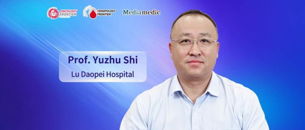
Editor's Note: On August 21, 2026, the 14th Lu Daopei Hematology Forum officially opened at Hebei Yanda Lu Daopei Hospital. Over the past 14 years, the conference has established a strong academic foundation. This year's meeting focused on cutting-edge topics including hematopoietic stem cell transplantation, cellular immunotherapy, and precision diagnosis and treatment of hematologic diseases, bringing together experts and scholars from across China to discuss advances in the field. During the conference, Oncology Frontier – Hematology Frontier invited Professor Yuzhu Shi from Lu Daopei Hospital for an interview. He shared his insights into the pathogen characteristics of central nervous system (CNS) infections after hematopoietic stem cell transplantation (HSCT), key imaging features for differential diagnosis, and the clinical value and future development of imaging technologies.Oncology Frontier – Hematology Frontier: Your presentation at this conference focused on “Imaging Features of Infectious Complications of the Central Nervous System After HSCT.” Could you introduce the incidence of CNS infections after HSCT and the major types of pathogens involved? What role does imaging play in early recognition and diagnosis?
Professor Yuzhu Shi: CNS infections following hematopoietic stem cell transplantation can be caused by a wide range of pathogens, including viruses, bacteria, and fungi, as well as less common pathogens such as Toxoplasma gondii and Mycobacterium tuberculosis. Clinically, fungal infections are the most common, followed by bacterial and viral infections, while tuberculosis and toxoplasmosis are relatively rare. In recent years, as the number of HSCT procedures has increased, the incidence of viral CNS infections has gradually risen.
Different pathogens cause distinct pathological changes and consequently produce different imaging manifestations. In the early stages of disease, when specific diagnostic methods may be limited, imaging is a key tool for establishing a timely diagnosis once patients develop relevant clinical symptoms. It therefore plays a critical role in the early identification of CNS infections.
Oncology Frontier – Hematology Frontier: What are the characteristic imaging manifestations of different CNS pathogens, such as fungi, viruses, and bacteria? How can imaging features be used for differential diagnosis?
Professor Yuzhu Shi: Bacterial and fungal infections are the most common in clinical practice. Because different pathogens have fundamentally different routes of infection and pathological changes, their imaging characteristics also differ and can be used for differential diagnosis.
In terms of the route of infection, bacterial infections commonly enter the cranial cavity through hematogenous dissemination and are often accompanied by pulmonary infection, such as infections caused by Legionella or Nocardia. Fungal infections, particularly those caused by Mucorales, Aspergillus, and Fusarium, often invade the cranial cavity directly from the paranasal sinuses and are frequently accompanied by sinus infection. Candida and certain other fungi may also disseminate to the CNS through the bloodstream. Viral infections are relatively less common clinically.
From the perspective of key imaging findings, bacterial infections are primarily characterized by abscess formation. On diffusion-weighted imaging (DWI), abscesses typically show high signal intensity with restricted diffusion. In contrast, fungal infections are mainly characterized by coagulative necrosis, and diffusion is generally not restricted, resulting in relatively low signal intensity on DWI. This difference represents one of the most important imaging features for distinguishing fungal from bacterial infections.
Viral infections generally do not form abscess cavities or solid mass lesions. Instead, they tend to cause diffuse abnormalities and may be accompanied by cytotoxic cerebral edema, which can also appear as high signal intensity on DWI. Lesions frequently involve the limbic system, with typical involvement of the hippocampus and temporal lobes, and patients often present clinically with seizures. CNS infections caused by tuberculosis or Toxoplasma gondii are relatively uncommon.
Oncology Frontier – Hematology Frontier: What additional potential does imaging have for early screening and treatment response assessment of CNS infections after HSCT? What do you see as the future directions of this field?
Professor Yuzhu Shi: For CNS infections following HSCT, CT can be used for initial screening. When intracranial fungal, bacterial, or other infections are strongly suspected, MRI is generally recommended as the preferred imaging modality. In addition to conventional MRI sequences, diffusion-weighted imaging is recommended because it is particularly valuable for differentiating fungal from bacterial infections. Susceptibility-weighted imaging (SWI) and magnetic resonance spectroscopy (MRS) can also be incorporated to further assist in differentiating abscesses from other primary lesions and improve diagnostic accuracy.
With continued advances in imaging technology, the combined application of more specialized imaging sequences is expected to further improve the early differential diagnosis of CNS infections and enhance the clinical value of imaging in early screening and treatment response assessment.
Expert Profile

Beijing Lu Daopei Hospital
Beijing Lu Daopei Hematology Hospital
Director, Department of Radiology, Hebei Yanda Lu Daopei Hospital
Committee Member, Third Committee of the Radiology Professional Committee, China Association of Non-Public Medical Institutions
Committee Member, Third Committee of the Hematology Professional Committee, China Association of Non-Public Medical Institutions
Committee Member, Cellular Immunotherapy Professional Committee, China Health Science and Technology Promotion Association
Committee Member, Pediatric Radiology Group, 13th Committee of the Radiology Branch of the Beijing Medical Association
Committee Member, Third Radiology Professional Committee, Beijing Association of Non-Public Medical Institutions
Committee Member, Second Hematologic and Lymphoid Tumors Professional Committee, Beijing Anti-Cancer Association
Since September 2011, Professor Yuzhu Shi has worked in the radiology departments of Beijing Lu Daopei Hospital, Beijing Lu Daopei Hematology Hospital, and Hebei Yanda Lu Daopei Hospital, specializing in imaging diagnosis related to complications following hematopoietic stem cell transplantation. During this period, he has published dozens of articles on post-HSCT imaging findings in Chinese core journals and SCI-indexed journals.
He has also performed CT-guided biopsy procedures in more than 100 post-HSCT patients, involving the lungs, superficial lymph nodes, mediastinal masses, liver, spleen, pancreas, bone, and other sites. Through this work, he has accumulated extensive clinical experience and provided valuable support for clinical diagnosis and treatment.