

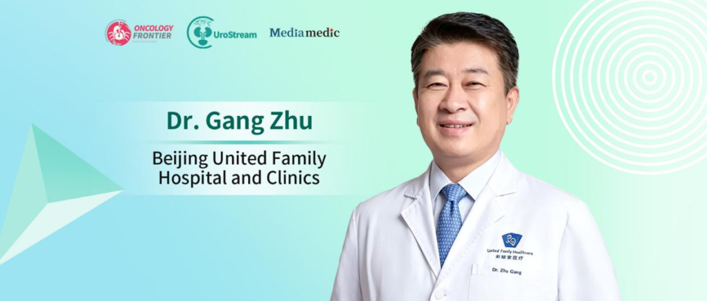
Editor's Note: The 2026 Pujiang Urological Oncology Conference was held in Shanghai, bringing together experts from China and abroad to discuss standardized diagnosis and treatment, innovations in surgical techniques, and advances in precision medicine in urologic oncology. At this year's conference, the application of artificial intelligence (AI) and holographic imaging (three-dimensional reconstruction) in robotic surgery emerged as a major focus. Oncology Frontier – UroStream invited Professor Gang Zhu of Beijing United Family Hospital for an interview to discuss the clinical value of AI and holographic imaging, strategies for functional preservation, and the translational challenges involved in moving innovative technologies from demonstration to routine clinical practice.Integrating Holographic Imaging with Robotic Platforms to Enhance Preoperative Planning and Intraoperative Identification
01
With the development of AI and holographic imaging technologies, urologic oncology surgery is gradually moving beyond procedures based solely on two-dimensional imaging toward more three-dimensional and individualized preoperative planning. In your view, what are the main practical benefits of these technologies for robotic surgery today?
Professor Gang Zhu: First, I would like to thank Professor Dingwei Ye and his team for inviting me to participate in the 2026 Pujiang Urological Oncology Conference. It is also my great pleasure to accept this interview with Oncology Frontier.
Earlier this month, we attended the “New Horizon Global Medical Forum” in Hong Kong, where one session specifically focused on geriatric medicine and longevity medicine. According to relevant projections, China’s average life expectancy is expected to reach 80 years by 2030, while human lifespan may have the potential to increase further. Achieving healthy longevity requires multidisciplinary efforts, with cancer prevention and treatment being a crucial component.
In recent years, more precise robotic surgery, increasingly effective systemic therapies, and innovative approaches such as mRNA cancer vaccines have continued to advance. These developments are helping us control tumors more safely and effectively, enabling patients not only to “live longer” but also to “live better.”
Coming back to your question, the application of holographic imaging in robotic surgery essentially addresses a longstanding core challenge in surgery: how to achieve more precise tumor resection while preserving normal tissues and organ function to the greatest extent possible.
By using AI to transform conventional two-dimensional imaging into three-dimensional visual models through three-dimensional reconstruction, holographic imaging can assist surgeons with preoperative planning and intraoperative navigation. When integrated with robotic platforms, it has the potential to further improve tumor localization, anatomical identification, and precision resection.
Therefore, the integration of holographic imaging with robotic surgery represents an important pathway for addressing complex clinical challenges and is likely to become an important direction in the future development of urologic oncology surgery.
From Tumor Resection to Functional Preservation: Toward More Individualized Treatment Goals
02
Urologic oncology surgery aims not only to achieve tumor control but also increasingly emphasizes long-term outcomes such as renal function, urinary continence, and sexual function. How can AI and image-guided navigation help surgeons transition from simply “removing the tumor” to achieving precise resection while maximizing functional preservation?
Professor Gang Zhu: Looking back at the development of oncologic surgery, early surgical approaches often prioritized maximal tumor removal, sometimes at the cost of sacrificing an entire organ or critical anatomical structures. This led to extensive or “en bloc” resection strategies.
With advances in medicine, imaging, and clinical research, we have gradually recognized that for certain localized tumors, local resection or tumor enucleation can also achieve favorable oncologic control.
This concept has gained increasing support in urology. Take renal tumors as an example. For appropriately selected patients, nephron-sparing surgery can achieve oncologic control comparable to radical nephrectomy while preserving renal function more effectively and reducing the long-term risks of chronic kidney disease and related metabolic complications.
This trend is not limited to renal tumors. In prostate cancer, for example, patients differ substantially in terms of age, fertility needs, and functional preservation priorities.
We once treated a 50-year-old patient with prostate cancer who had not yet had children. He hoped to control his cancer while preserving fertility, sexual function, and urinary continence to the greatest extent possible. After a comprehensive evaluation, we performed focal ablation using irreversible electroporation (IRE). Following treatment, these functions were all relatively well preserved.
This case illustrates that treatment decisions should not focus solely on the tumor. We also need to take the patient’s life stage and individual needs fully into account.
As an auxiliary decision-making tool, holographic imaging can provide support during diagnosis, preoperative planning, and treatment, helping us develop more precise strategies that better align with individual patient needs.
The transition from “removing the organ” to “preserving function” represents not only technological progress but also a profound shift in treatment philosophy. AI and image-guided navigation are becoming important forces driving this transformation.
Bringing AI into Routine Clinical Practice: High-Quality Inputs, Physician Verification, and Clear Accountability Are All Essential
03
As AI-assisted surgery moves from technological demonstrations toward routine clinical application, challenges remain in areas such as standardization, data quality, learning curves, and clinical validation. What do you believe are the most important barriers to overcome in the next stage to ensure that these innovations genuinely transform everyday surgical decision-making?
Professor Gang Zhu: This is a critical issue that must be addressed in clinical translation. AI is rapidly entering both everyday life and healthcare, and clinicians are increasingly recognizing its value and becoming more willing to use it.
At United Family Hospital, for example, we developed the “UFH AI” system relatively early. With the patient’s informed consent, physicians can use AI to assist in recording clinical conversations during natural interactions with patients. The system can then generate information such as the chief complaint, history of present illness, past medical history, medication history, diagnosis, and treatment plan in accordance with medical documentation standards. It can also provide diagnostic and treatment suggestions for physicians’ reference.
This allows physicians to devote more time to observing patients and engaging in meaningful human communication. As mentioned earlier, holographic imaging is also generated using AI technology. It is foreseeable that AI will have an extremely broad range of applications in clinical medicine.
At the same time, however, two core issues must receive particular attention.
First, accountability must be clearly defined, and we must remain alert to AI “hallucinations.” AI may generate inaccurate or even erroneous information. Although such problems are becoming less frequent as the technology evolves, we must remain fully aware that, at the current stage, physicians remain ultimately responsible for clinical decision-making. When using AI, doctors must rigorously verify its outputs and incorporate them into clinical decision-making only after confirming that the information is accurate and applicable. AI can provide assistance, but it cannot replace the professional judgment of clinicians.
Second, we must ensure high-quality data at the source and adhere to the principle of “high-quality evidence in.” Only when AI systems are built upon reliable and authoritative domestic and international clinical guidelines and evidence-based medical literature, together with rigorous data governance and updating mechanisms, can they generate more trustworthy outputs.
The principle of “garbage in, garbage out” reminds us that we must focus on data quality and system design from the outset. By grounding AI applications in high-level evidence, we can ensure that these technologies genuinely help clinicians make more accurate and reliable diagnostic and treatment decisions.
Summary
Professor Gang Zhu’s insights provide a clear picture of the evolving role of AI and holographic imaging in urologic oncology surgery. From preoperative holographic planning to precise intraoperative identification, and from radical tumor control to functional preservation, intelligent technologies are reshaping the paradigm of surgical practice.
At the same time, the faster these technologies develop, the more important it becomes to uphold the principle that physicians remain the primary decision-makers and evidence-based medicine remains the foundation. Only by maintaining high standards for data quality, clinical verification, and accountability can technological innovation be successfully translated into meaningful benefits for patients.
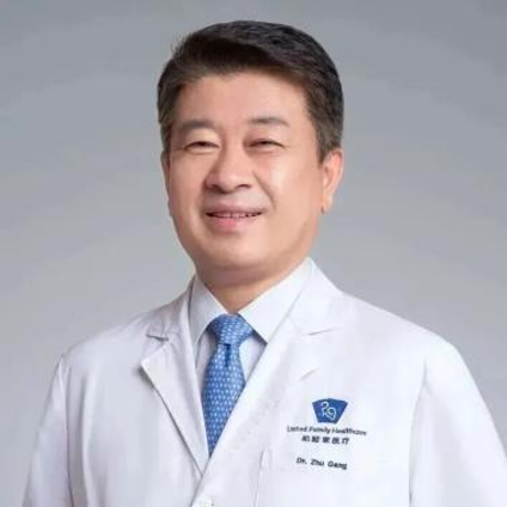
Professor Gang Zhu