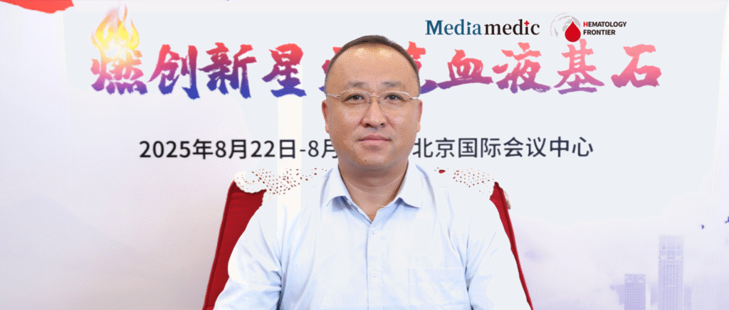
On August 22–23, 2025, the 13th Lu Daopei Hematology Conference was held in Beijing, jointly organized by the Beijing Health Promotion Association and the Guangzhou Kapok Oncology and Rare Disease Foundation, and hosted by the Beijing Lu Daopei Hematology Institute. The conference gathered world-leading hematology experts and focused on hematopoietic stem cell transplantation, cellular therapy, and precision treatment of hematologic malignancies. With over a thousand participants, the event delivered a high-level, in-depth academic exchange. During the meeting, Professor Yuzhu Shi from Beijing Lu Daopei Hospital presented a keynote lecture on the clinical value of abdominal CT in evaluating abdominal complications following hematopoietic stem cell transplantation (HSCT). Oncology Frontier – Hematology Frontier invited Professor Shi to provide further insights into this important topic, offering practical guidance and theoretical reference for optimizing transplant management strategies.PART 1
Abdominal complications are a major challenge in post-HSCT management. Compared with other imaging modalities or laboratory tests, what do you consider the unique advantages and core clinical value of abdominal CT in evaluating such complications?
Professor Yuzhu Shi: Abdominal CT is fast, convenient, and noninvasive, making it highly valuable in assessing abdominal complications after HSCT. Some complications can be directly diagnosed by CT, while others require correlation with clinical presentation and laboratory findings for a comprehensive evaluation.
In addition, CT can support differential diagnosis of related conditions. These advantages underscore the important role of abdominal CT in the diagnosis and management of abdominal complications following transplantation.
PART 2
From the perspective of hematologists, what are some of the unique challenges in interpreting abdominal CT images in post-HSCT patients? Could you share several CT features that are particularly suggestive or useful for differential diagnosis in this setting?
Professor Yuzhu Shi: Post-transplant abdominal CT findings can be complex, and in many cases radiologists need to collaborate closely with clinicians to aid in interpretation and guide diagnostic direction.
For example, hepatic sinusoidal obstruction syndrome (SOS) can be clearly diagnosed on CT. Other findings, though relatively uncommon, are diagnostically significant. These include the “target sign” of the bowel, bowel wall thickening, or intramural gas—subtle changes often difficult to detect through clinical examination but identifiable with imaging. Such features not only help determine the etiology but also allow evaluation of treatment response.
Compared with invasive procedures such as endoscopy, abdominal CT offers distinct advantages: it does not require invasive intervention, has broad indications, is quick and simple to perform, and carries minimal risk of procedure-related complications. These features make CT an especially valuable tool in both diagnosis and follow-up of post-transplant abdominal complications.
PART 3
Given current clinical needs, what future directions should research on abdominal CT in post-HSCT complications focus on? Which are most likely to translate into breakthroughs in clinical practice?
Professor Yuzhu Shi: Abdominal CT is particularly valuable in the evaluation of gastrointestinal complications after HSCT, including differentiation of infectious lesions.
It also plays an important role in assessing gastrointestinal involvement of graft-versus-host disease (GVHD) and monitoring treatment response. Unlike endoscopy, which can usually be performed only once or twice during therapy, abdominal CT can be repeated multiple times, enabling dynamic observation of disease progression.
By integrating imaging findings with clinical data, CT can support disease staging, response assessment, and identification of acute abdominal events, thus providing essential diagnostic information to guide clinical decision-making.
Expert Profile

Professor Yuzhu Shi Beijing Lu Daopei Hospital Beijing Lu Daopei Hematology Hospital Director of Radiology, Hebei Yanda Lu Daopei Hospital
- Member, 2nd Committee of the Radiology Professional Committee, China Non-Public Medical Institutions Association
- Member, 2nd Committee of the Hematology Professional Committee, China Non-Public Medical Institutions Association
- Member, Cellular Immunotherapy Committee, China Association for the Promotion of Human Health Science and Technology
- Member, Pediatric Group, 13th Committee of the Radiology Branch, Beijing Medical Association
- Member, 2nd Radiology Committee, Beijing Non-Public Medical Institutions Association
Professor Shi graduated in July 2002 with a bachelor’s degree in Medical Imaging from Mudanjiang Medical College and has been a member of the Chinese Communist Party since her student days. She began her career in the Department of Radiology at the First Affiliated Hospital of Henan University, where she received extensive training and accumulated significant diagnostic experience. Since September 2011, she has worked in the Radiology Departments of Beijing Lu Daopei Hospital and Hebei Yanda Lu Daopei Hospital, specializing in imaging diagnosis related to hematopoietic stem cell transplantation.
She has published dozens of papers on imaging manifestations after transplantation in leading Chinese core journals and SCI-indexed journals. She has also completed nearly one hundred CT-guided biopsies in post-HSCT patients involving the lung, superficial lymph nodes, mediastinal masses, liver, spleen, pancreas, and bone tissue, providing crucial diagnostic and therapeutic support. Her extensive experience has significantly contributed to improving post-transplant patient care.

