
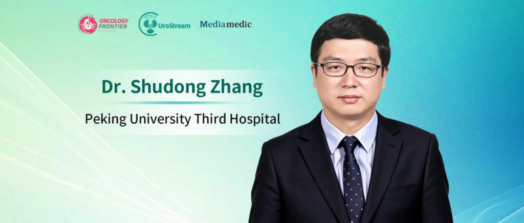
Editor's Note: The 2026 Pujiang Urological Oncology Academic Conference was held in Shanghai, bringing together experts in urological oncology from China and abroad to discuss standardized diagnosis and treatment, surgical innovation, and advances in precision medicine. Upper tract urothelial carcinoma (UTUC) complicated by inferior vena cava (IVC) tumor thrombus is relatively rare and is associated with substantially increased surgical complexity and perioperative risk. Oncology Frontier · UroStream invited Professor Shudong Zhang of Peking University Third Hospital for an interview, during which he shared his team's experience and clinical perspectives on preoperative imaging assessment and risk stratification, the application of robotic surgery, and multidisciplinary team (MDT) management throughout the treatment journey.Multimodal Imaging and Precise Risk Stratification: Establishing a Foundation for Safe High-Risk Surgery
01
Oncology Frontier · UroStream: UTUC complicated by IVC tumor thrombus is relatively uncommon, and its presence significantly increases surgical complexity and perioperative risk. What key imaging assessments and risk stratification should be completed before surgery in these patients?
Professor Shudong Zhang: Patients with UTUC complicated by IVC tumor thrombus are at high risk, and the thrombus may also become detached. Therefore, the first priority of imaging assessment is to comprehensively define the extent of the tumor thrombus, including its upper and lower boundaries, location, morphology, and mobility; determine its relationship with the IVC wall; and identify whether concomitant bland thrombus is present.
First, multiparametric MRI of the IVC should be performed. Conventional imaging may not always adequately cover the right atrium above the thrombus and the venous segments below it, potentially compromising the overall assessment. Following repeated discussions with our radiology colleagues, we developed a dedicated multiparametric IVC MRI protocol. Through multi-sequence and multiplanar imaging, we aim to visualize the tumor thrombus and relevant vascular anatomy as comprehensively as possible, providing a basis for determining thrombus level, extent, and venous wall involvement.
Second, comprehensive systemic staging should be completed. PET-CT is not mandatory for every patient, but it can provide a more comprehensive assessment of overall disease burden. Because UTUC complicated by venous tumor thrombus carries a relatively high risk of lymph node metastasis, regional lymph nodes, adjacent organs, and major organs such as the liver and lungs should also be systematically evaluated for metastatic disease.
Third, transesophageal echocardiography (TEE) should be given particular attention. For high-level tumor thrombi, especially those extending toward the suprahepatic IVC or right atrium, TEE can help assess the upper extent and mobility of the thrombus as well as the risk of detachment. It can also provide real-time intraoperative monitoring of the thrombus and thrombectomy process, supporting anesthetic and surgical safety.
Risk stratification should not be based solely on thrombus level. Factors including thrombus width, size, morphology, consistency, venous wall involvement, and concomitant bland thrombus should all be considered. Our team is also incorporating AI and radiomics to develop a more refined scoring system, with the goal of providing quantitative support for predicting surgical complexity and selecting treatment pathways.
Tailoring the Surgical Approach According to Thrombus Level and Vascular Involvement
02
Oncology Frontier · UroStream: How should an individualized surgical approach be designed according to the extent of tumor thrombus, degree of vascular involvement, and characteristics of the primary tumor? What role can robotic or minimally invasive techniques play in this challenging setting?
Professor Shudong Zhang: The level, morphology, consistency, and relationship of the tumor thrombus with the venous wall all influence surgical strategy. Therefore, a detailed and individualized surgical plan is essential. Based on preoperative imaging and risk stratification, we use imaging-processing software to construct three-dimensional models. By viewing the spatial relationships between the tumor thrombus, IVC, renal hilar vessels, and surrounding critical organs from multiple angles, and following MDT discussion, we determine the appropriate surgical approach.
For infradiaphragmatic or infrahepatic tumor thrombi, the sequence of vascular clamping can be planned according to the upper and lower boundaries of the thrombus and the status of collateral circulation, followed by thrombectomy and vascular repair.
For suprahepatic tumor thrombi or those extending into the right atrium, the surgical approach must be further tailored according to whether the thrombus is adherent to or invading the venous wall and the risk of detachment. Anesthesia, ultrasound monitoring, and, when necessary, circulatory support strategies should also be planned in advance.
Robotic surgery offers advantages in deep surgical field exposure, lymph node dissection, identification of critical structures, precise suturing, and vascular management, thereby facilitating minimally invasive approaches for complex tumor thrombus surgery. However, its value must be assessed objectively: not every patient is suitable for robotic surgery. Patient selection must be matched to the thrombus classification, degree of vascular wall involvement, surgical team’s experience, and capacity for emergency management.
At our center, we have performed more than 800 surgeries for various types of venous tumor thrombi, including more than 400 robotic procedures. Over the past five years, robotic surgery has accounted for more than 85% of cases. These experiences indicate that robotic technology can be an important driver of minimally invasive surgery for complex tumor thrombi. However, high-quality imaging assessment, standardized workflows, and an experienced multidisciplinary team remain the foundation for safe implementation.
From Perioperative Collaboration to Systemic Therapy: Establishing Comprehensive MDT Management
03
Oncology Frontier · UroStream: For these high-risk UTUC patients, high-quality surgery alone may still be insufficient to improve long-term outcomes. Should the ideal treatment model further integrate systemic therapy with surgery? How should multidisciplinary teams become involved earlier?
Professor Shudong Zhang: MDT management is essential for challenging and high-risk urological malignancies. Multidisciplinary collaboration should not begin only when difficulties arise during surgery. Instead, it should be initiated at the first evaluation and continue throughout diagnosis, perioperative treatment, postoperative systemic therapy, rehabilitation, and follow-up.
First, MDT involvement should be moved forward to the diagnostic and preoperative decision-making stages. Before surgery, the pathology and radiology teams should establish the diagnosis and stage as accurately as possible. Urologists, anesthesiologists, echocardiography specialists, cardiologists, pulmonologists, and interventional vascular specialists should jointly assess the risks of tumor thrombus detachment, pulmonary embolism, hemodynamic instability, and major vascular management. For patients with pulmonary embolism or complex vascular conditions, the relevant teams should formulate contingency plans in advance.
Second, postoperative systemic therapy and rehabilitation should be closely integrated. Surgery represents only one component of the overall treatment pathway. After surgery, systemic treatment and follow-up should be jointly planned by medical oncology, radiation oncology, and relevant specialists based on pathological stage, lymph node status, physical recovery, and recurrence risk. Rehabilitation, nutritional support, and complication management should also be incorporated. Only by systematically integrating surgery with systemic therapy can we further improve long-term outcomes.
Third, specialized subspecialty teams should drive comprehensive, refined management. Because these cases are rare and technically demanding, large-volume centers should establish dedicated subspecialty teams and standardized MDT workflows covering diagnosis, treatment, and follow-up to ensure continuous patient benefit throughout the entire treatment journey.
Summary
Professor Shudong Zhang provided a comprehensive overview of the precision surgical strategy for UTUC complicated by IVC tumor thrombus—from multimodal preoperative assessment using multiparametric IVC MRI, PET-CT, and transesophageal echocardiography, to AI- and radiomics-based risk stratification, and from three-dimensional model-guided individualized surgical planning to robotic minimally invasive thrombectomy. Ultimately, the strategy centers on comprehensive MDT management spanning the preoperative, intraoperative, and postoperative stages.
For these high-risk patients, surgery is only one component of the overall treatment strategy. Closer integration of systemic and surgical therapy, together with early multidisciplinary involvement, is key to improving long-term outcomes.
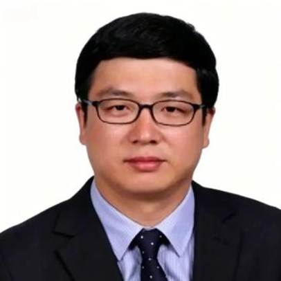
Professor Shudong Zhang
Beijing Peking University Third Hospital