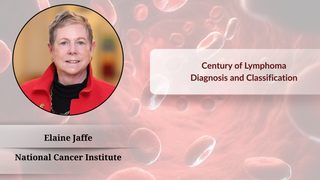
Editor's Note: The International Hematologic Oncology Pathology Conference brought together top experts in hematopathology research from home and abroad for in-depth discussions on cutting-edge topics such as lymphoma diagnosis, classification evolution, and disease discovery methods. This article is based on the insightful report "The Journey of Disease Discovery: A Personal Voyage" by Professor Elaine Jaffe, an internationally renowned hematopathology expert from the National Cancer Institute (NCI), systematically reviewing the unique value of the microscope in disease discovery, the evolution of lymphoma classification systems, and sharing unique insights into frontier research on follicular lymphoma, with the aim of providing reference for clinical and scientific colleagues.The Microscope: An Enduring Tool for Disease Discovery and Rudolf Virchow’s Legacy
In today’s rapidly advancing modern medicine, despite the rapid development of molecular biology techniques, the microscope, as a fundamental tool for disease discovery, still holds undeniable core value. Professor Elaine Jaffe emphasized in her report that Rudolf Virchow, known as the father of surgical pathology, first proposed the theory of cellular disease and coined terms like “lymphosarcoma” and “leukemia” over a hundred years ago, laying the foundation for disease research. Professor Jaffe pointed out that although there are many advanced tools for diagnosing lymphoma today, the microscope still holds a place in our diagnostic toolkit and serves as the starting point for disease discovery and understanding.

Early Exploration and Challenges in Lymphoma Classification Systems
Professor Jaffe reviewed the early history of lymphoma classification. She mentioned that when she first entered the field of pathology, the Rappaport system was the main basis for lymphoma classification. This system purely described tumors as lymphocytic or histiocytic based on cell size, and distinguished them by nodular or diffuse morphology, but did not presuppose a connection between nodular lymphoma and lymphoid follicles.

Around the same time, Professor Karl Lennert began his pioneering research on the lymphatic system, also relying solely on microscopic observation. Through in-depth study of normal germinal centers, he identified follicular dendritic cells, small lymphocytes, and large lymphocytes, and observed that these cell types also existed in the disease then known as nodular lymphoma. Professor Lennert therefore proposed that this disease indeed originated from normal germinal centers, laying the foundation for subsequent classification innovations.

The 1970s Revolution: Immunological Advances Drive Leaps in Lymphoma Diagnosis
The 1970s witnessed a true revolution in lymphoma diagnosis. Professor Jaffe shared that significant progress was made in lymphoma and leukemia treatment during that time, accompanied by breakthrough understanding in immunology, leading to new insights into the nature of immune cells. More importantly, new techniques capable of characterizing lymphocytes and histiocytes began to emerge.

During her postdoctoral fellowship, Professor Jaffe collaborated with immunologists at the National Institute of Allergy and Infectious Diseases (NIAID) to study lymphocytes in vitro. They discovered that B cells possess C3D receptors (now known as CD21), which could be detected in cell suspensions by red blood cells coated with antibodies and complement (EAC). Professor Jaffe cleverly used the “hanging drop technique” to layer EAC red blood cells onto frozen sections, observing their binding to reactive germinal centers in normal lymph nodes. Subsequently, she applied this method to cases of nodular lymphoma, observing the same phenomenon. This research powerfully demonstrated that nodular lymphoma indeed originated from follicular B lymphocytes, providing important evidence for the immunological origin of lymphoma. This study was eventually published in the New England Journal of Medicine and became a classic citation.

The 1970s saw the introduction of many new techniques for identifying T cells and B cells, such as sheep red blood cells for detecting CD2 and surface immunoglobulin for B cells. Along with this new knowledge, there was keen interest in developing lymphoma classifications that more closely correlated with the immune system. However, despite six classification schemes being proposed and discussed at international meetings, pathologists could not reach a consensus. To address this, the National Cancer Institute (NCI) proposed the “Working Formulation for Clinical Application,” essentially a terminological refinement of the Rappaport system. This classification failed to clearly define disease entities, and most categories, such as “diffuse mixed type,” remained highly heterogeneous, relying solely on H&E morphology and failing to fully utilize the emerging techniques of the time. Although accepted in the United States, the Kiel system was more popular in Europe and Asia.


The 1980s-90s Milestones: The Rise of Immunohistochemistry and Molecular Genetics
Over the subsequent ten to twenty years, rapid progress was made in characterizing lymphoproliferative disorders. Clive Taylor and David Mason developed immunohistochemical techniques usable on paraffin-embedded sections, eliminating the need for frozen sections. Köhler and Milstein’s monoclonal antibody technology revolutionized diagnosis, and David Mason also began developing monoclonal antibodies for use in paraffin sections. Immunoglobulin gene rearrangement was proven to be a lineage and clonality marker. Della Favera identified the translocated c-MYC gene in Burkitt lymphoma, and subsequently, Croce and Sugimoto identified BCL2 as a key gene in follicular lymphoma. These breakthrough advances provided unprecedented tools for the precise diagnosis of lymphoma.


REAL Classification: A New Paradigm for Lymphoma Classification
Based on this new knowledge, Peter Isaacson, in collaboration with Harold Stein, believed that they could move beyond the Working Formulation. He convened about 20 hematopathology experts from around the world in London to discuss three topics: the relationship between intermediate lymphoma and mantle cell lymphoma (confirming they were the same disease and related to the mantle zone), Peter Isaacson’s concept of MALT lymphoma (related to gastrointestinal and mucosa-associated lymphomas, which were not recognized by existing classifications at the time), and the association between Hodgkin’s disease and non-Hodgkin’s lymphoma (both possibly originating from B lymphocytes).
At the first meeting of the International Lymphoma Study Group, experts reached a consensus on several issues, realizing that they were indeed speaking the same language. As Nancy Harris wrote in the REAL classification manuscript: “We believe that the most practical approach to classifying lymphomas at present is simply to define the disease entities we believe can be identified by currently available morphological, immunological, and genetic techniques.” This proposal was published in Blood.
The REAL (Revised European-American Lymphoma) classification represented a new paradigm for classifying lymphoproliferative disorders. It defined disease entities based on clinical and laboratory features (including clinical presentation and course), and recognized that the site of involvement often served as a marker of underlying biological differences (e.g., MALT lymphoma). This classification was built on the consensus of 19 experts, not a theoretical scheme, but based on published data, with only entities supported by at least two peer-reviewed references being included. The REAL classification was later validated in a large international study led by Jim Armitage, and its disease-oriented approach offered numerous advantages in lymphoma classification, being crucial for developing new therapies and studying the pathogenic mechanisms of specific diseases.
WHO Classification: A Product of Global Collaboration and Consensus
The REAL classification naturally became the basis for the subsequent WHO classification. In 1993-1994, Les Sobin invited the Society for Hematopathology to develop a classification for the third edition of the WHO Blue Book. After Paul Kleihues took over as IARC Director in 1994, he transformed the Blue Book series into its current format, integrating pathological, genetic, and clinical findings, which aligned perfectly with the philosophy of the REAL classification.
Another important step in the development of the WHO classification was the realization of the need for a Clinical Advisory Committee (CAC) meeting, and that clinicians must be involved in the process. The CAC was jointly organized by the Society for Hematopathology and EHP, with 200 invited participants from 24 countries. Clinicians and pathologists collaboratively discussed various disease entities, and the meeting reports were published in clinical and pathological journals, and importantly, this occurred before the final draft of the WHO Blue Book was finalized.
Professor Elaine Jaffe pointed out that compared to the 1975 meeting, the 1997 meeting was able to achieve consensus because the classification was a collective effort of multiple experts, with pathologists and clinicians working side-by-side. At that time, we had a better understanding of the normal immune system, paraffin immunohistochemistry could be used to detect most relevant antigens in routine biopsies, and molecular studies could also be performed on paraffin-embedded tissue to identify clonal and genetic features. Most importantly, these proposals could be empirically tested. This model was successfully applied to the production of the third, fourth, and subsequent WHO Blue Books.
Cornerstone and Evolution of Lymphoma Disease Discovery
Professor Jaffe re-emphasized that the microscope is an important tool for disease discovery. All diseases she presented were first identified through conventional pathology, histology, and clinical criteria, and careful descriptions of these disease entities in turn promoted understanding of their pathogenesis. She admitted that today, molecular biology is becoming increasingly important in diagnosis, especially in diffuse large B-cell lymphoma, where molecular biology may be even more crucial than the microscope. Image: Lymphoma morphology under the microscope.
Subsequently, Professor Jaffe shared two research cases on follicular lymphoma. Early Oncogenesis and Clonal Evolution: Follicular Lymphoma in situ In 1992, Professor Jaffe received a consultation case: a 23-year-old female presented with an isolated enlarged inguinal lymph node. Pathologists found a small number of cells with a T(14;18) translocation. Professor Jaffe used the BCL2 monoclonal antibody provided by David Mason to stain tissue sections and found that BCL2-positive cells were selectively located within residual reactive germinal centers, indicating early involvement by follicular lymphoma. After 15 years of follow-up, the patient showed no evidence of follicular lymphoma.
In the following years, they encountered 23 similar cases, which they termed “Follicular Lymphoma in situ.” These lymph nodes initially appeared reactive, with well-structured germinal centers, but if stained with BCL2 antibody, BCL2-positive cells were found within the follicles, suggesting early involvement by follicular lymphoma. They demonstrated that these BCL2-positive cells were clonal by PCR and contained BCL2 gene rearrangements. Subsequent studies showed that unless incidentally discovered during staging, most patients never developed follicular lymphoma, with a risk of less than 3%.
Concurrently, Bertrand Nadel and his team found that BCL2 gene rearrangement is very common in healthy individuals, reaching as high as 70% in adults over 50, and increasing with age and pesticide use by farmers. These BCL2-rearranged cells are not naive B cells, but rather class-switched memory B cells that have undergone germinal center reactions. Follicular lymphoma in situ and follicular lymphoma-like B cells in the blood are part of the same process and may coexist in the same patient.


By microdissecting BCL2-positive cells from follicular lymphoma in situ, partially involved lymph nodes, duodenal-type follicular lymphoma, and classic follicular lymphoma, Professor Jaffe’s laboratory found that in situ lesions and duodenal-type follicular lymphoma had very few genomic alterations, while classic follicular lymphoma showed higher levels of genomic alterations. The study showed that follicular lymphoma in situ not only has BCL2 gene rearrangement but also other genomic lesions associated with follicular lymphoma, often positive for CREBBP, TNFSF14, and EZH2 mutations. This provided a conceptual model for the pathogenesis and development of follicular lymphoma and revealed the possibility of recurrence through linear and divergent evolution, and even transformation to other phenotypes or lineages.
Lineage Plasticity and Transformation: Secondary Histiocytic and Dendritic Cell Sarcomas
Professor Jaffe presented a second case: a patient with follicular lymphoma who developed a highly anaplastic and pleomorphic tumor three years after diagnosis. This tumor lacked all B-cell markers but expressed a histiocytic and dendritic cell phenotype. All 8 cases showed clonal identity with the previous follicular lymphoma through sequencing and BCL2 gene rearrangement and expressed transcription factors known to be associated with histiocytic differentiation. Notably, PAX5 was consistently negative, as were other B-cell lineage markers.
Previous mouse model studies had shown that blocking PAX5 could reprogram mature B cells into mature macrophages, and this may also occur in the human system. Their study further revealed that these secondary histiocytic sarcomas all acquired mutations in the RAS-RAF-MAP kinase pathway but retained many mutations from the preceding follicular lymphoma. The molecular mechanisms of PAX5 loss still need further elucidation.
Outlook and Conclusion
In her summary, Professor Elaine Jaffe emphasized that lymphoproliferative disorders are characteristics of the normal immune system, and they provide us with information for understanding normal and neoplastic cells. Lymphocytes, as part of their normal function, migrate or disseminate, and non-clonal lymphoproliferation is not confined locally but disseminated according to normal lymphocyte homing mechanisms. Therefore, for lymphoproliferative disorders, the distinction between benign and malignant is blurred. We talk about malignant lymphoma, but there is no benign lymphoma (or we call them other diseases, such as MALT lymphoma). Finally, Professor Jaffe concluded that the observations we make under the microscope and clinically are the starting point for disease discovery. Although molecular biology is increasingly important, the intuitive information provided by the microscope remains an indispensable foundation for a deeper understanding of disease pathogenesis and progression, guiding precise diagnosis and treatment strategies. This review is not only a personal journey but also a profound insight into the development history of the entire field of hematopathology. Through Professor Elaine Jaffe’s excellent presentation, the International Hematologic Oncology Pathology Conference once again highlighted the importance of integrating classic pathology with modern molecular techniques, providing valuable experience and insights for the global hematopathology community, and jointly promoting a new chapter in precise diagnosis and treatment of lymphoma. Provided/Interview Source: Oncology Outlook – Oncology News

