


Patients with advanced epidermal growth factor receptor (EGFR)-mutant non-small cell lung cancer (NSCLC) inevitably face the challenge of acquired resistance after first-line treatment with third-generation EGFR tyrosine kinase inhibitors (TKIs). Currently, there is no universally established standard of care following resistance. Options such as platinum-based chemotherapy, EGFR-TKI combination regimens, antibody–drug conjugates (ADCs), and novel targeted agents remain under investigation. For these patients, balancing antitumor activity, intracranial disease control, and long-term treatment tolerability remains a major clinical challenge.In this issue, we invited Professor Lijun Tian of the Affiliated Hospital of Shandong First Medical University to share a case involving a patient who had received multiple lines of treatment. The patient was a 52-year-old man with adenocarcinoma of the right lower lung, accompanied by multiple bilateral pulmonary metastases and mediastinal lymph node metastases at initial diagnosis. The clinical stage was cT2aN3M1, stage IVA. Molecular testing identified an EGFR exon 19 deletion (19-Del), while the programmed death-ligand 1 (PD-L1) tumor proportion score (TPS) and combined positive score (CPS) were both 0.
The patient received first-line aumolertinib, followed by dose-escalated furmonertinib combined with platinum-based chemotherapy, and subsequently sacituzumab tirumotecan. After further disease progression and the development of brain metastases, the patient received andaritini at 240 mg once daily (QD), an off-label treatment administered with the patient’s informed consent. Follow-up assessments at one and three months showed partial responses (PRs) in both the pulmonary lesions and intracranial metastases, with overall good tolerability.
Case Presentation
The patient was a 52-year-old man who was first admitted to our hospital on February 1, 2024, with the chief complaints of “throat discomfort for two months and a pulmonary mass detected one day earlier.”
History of Present Illness:
Approximately two months before admission, the patient developed persistent throat discomfort without an obvious precipitating factor. The nature of the discomfort was difficult to characterize, and during episodes he was unable to speak. He occasionally experienced coughing with a small amount of white sputum. He reported no fever, chills, chest tightness, or shortness of breath.
He had taken anti-inflammatory medication and Pangdahai (Sterculia lychnophora seeds), among other remedies, although the specific medications were unknown. These measures provided little relief. He subsequently attended the otolaryngology outpatient clinic at our hospital, where laryngoscopy revealed fixation of the right vocal cord and a polypoid lesion in the middle portion of the left vocal cord.
Chest computed tomography (CT) suggested lung cancer in the right lower lobe with multiple bilateral pulmonary metastases, as well as enlarged and mildly enlarged lymph nodes in both hilar regions and the mediastinum.
Physical Examination:
The ECOG performance status score was 1. No enlarged superficial lymph nodes were palpable. Breath sounds were clear bilaterally, with no wheezing or crackles. Cardiac rhythm was regular, heart sounds were strong, and no murmurs were heard over the cardiac valve areas. The abdomen was soft, with no palpable masses or abdominal tenderness. The liver was not palpable below the costal margin. No edema was present in either lower extremity.
Past Medical and Personal History:
The patient had a 30-year history of smoking, averaging 10 cigarettes per day, and a history of alcohol consumption lasting more than 30 years, with a daily alcohol intake of approximately 120 g.
Family History:
No significant family history was reported.
Marital and Reproductive History:
The patient married at age 25 and had one son.
Auxiliary Examinations:
Carcinoembryonic antigen (CEA): 71.40 ng/mL.
Chest CT (February 2, 2024):
A mass-like opacity was observed in the lateral basal segment of the right lower lobe, measuring approximately 4.0 × 3.2 cm on the largest cross-section. The lesion had an irregular shape and heterogeneous internal density, with areas of relatively high attenuation and cavitation. Its margins showed spiculation and lobulation, with adjacent pleural retraction. Some neighboring bronchi were narrowed or obstructed.
Multiple solid nodules were present in both lungs. The largest was located in the posterior segment of the right lower lobe and measured approximately 1.9 × 1.2 cm. Enlarged and mildly enlarged lymph nodes were also observed in both hilar regions and the mediastinum.
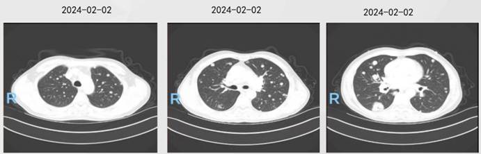
Baseline Chest CT (February 2, 2024)
Contrast-Enhanced Brain MRI (February 3, 2024): No evidence of brain metastases.
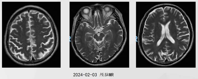
Baseline Brain MRI (February 3, 2024)
ECT (February 3, 2024): No significant abnormalities detected.
Ultrasound-Guided Percutaneous Biopsy of the Right Lung: Histopathological examination confirmed adenocarcinoma. PD-L1 TPS = 0%; CPS = 0.
PCR-Based Genetic Testing: (Right lung) EGFR exon 19 deletion (19-Del) mutation.
Clinical Diagnosis
- Adenocarcinoma of the right lower lobe of the lung (cT2aN3M1, stage IVA, EGFR exon 19 deletion mutation)
- Secondary malignant neoplasm of the mediastinal lymph nodes
- Secondary malignant neoplasms of both lungs
Treatment Course
First-Line Treatment: Aumolertinib Monotherapy
Starting February 6, 2024, the patient received oral aumolertinib 110 mg once daily (QD), together with oral rivaroxaban 10 mg QD. Chest CT performed after two months of treatment showed a partial response (PR). Follow-up assessments at four and eight months confirmed that the PR was maintained.
The patient developed a grade 1 rash during treatment but otherwise tolerated therapy well. Laboratory testing showed that the carcinoembryonic antigen (CEA) level had decreased to 21.40 ng/mL.
On January 6, 2025, routine follow-up examinations indicated progressive disease (PD). Progression-free survival (PFS) with first-line treatment was approximately 11 months.
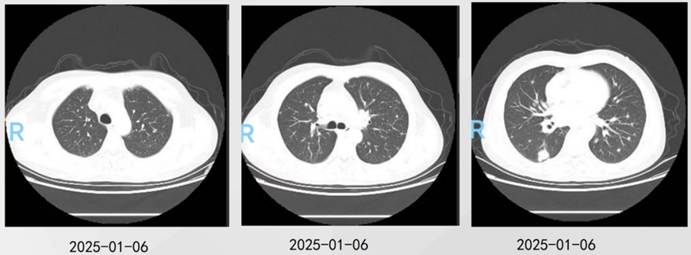
Chest CT on January 6, 2025, Indicating Disease Progression
Second-Line Treatment: Dose-Escalated Furmonertinib Combined with Platinum-Based Chemotherapy (Clinical Trial)
Following disease progression on January 7, 2025, a repeat biopsy was recommended, but the patient and family declined. Brain MRI and ECT showed no abnormalities.
The patient was enrolled in a clinical trial investigating high-dose furmonertinib monotherapy or furmonertinib combined with platinum-based chemotherapy for patients with locally advanced or metastatic NSCLC harboring sensitizing EGFR mutations whose disease had progressed following first-line treatment with a third-generation EGFR-TKI. Eligibility criteria included treatment with a third-generation EGFR-TKI for more than three months and a best response of PR.
Starting January 17, 2025, the patient received furmonertinib 160 mg orally once daily (QD), combined with pemetrexed disodium for injection and carboplatin injection every three weeks (Q3W). After six weeks, the response was assessed as PR, which remained the best overall response throughout treatment.
During treatment, the patient experienced grade 2 myelosuppression and grade 1 gastrointestinal adverse reactions.
On November 11, 2025, routine follow-up chest CT revealed progressive disease (PD). Progression-free survival (PFS) with second-line treatment was approximately 10 months.
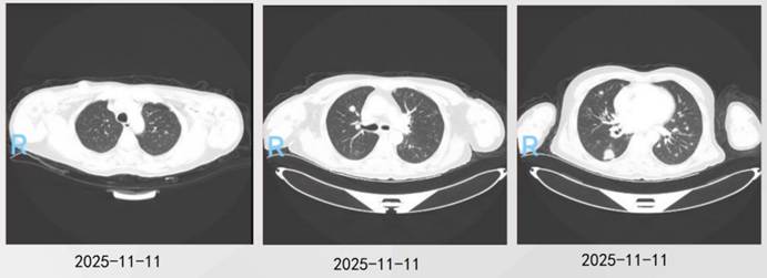
Chest CT on November 11, 2025, Indicating Disease Progression
Third-Line Treatment: Sacituzumab Tirumotecan (Clinical Trial)
Following disease progression on November 11, 2025, another biopsy was recommended, but the patient again declined. The patient had no symptoms suggestive of brain metastases, and ECT showed no significant abnormalities.
The patient was enrolled in a clinical trial comparing sacituzumab tirumotecan with docetaxel and received sacituzumab tirumotecan at 5 mg/kg every two weeks (Q2W).
Response assessments on November 15, 2025, and January 9, 2026, showed stable disease (SD). After 18 weeks of treatment, brain MRI on May 27, 2026, revealed newly developed brain metastases, and the response was assessed as progressive disease (PD).
During treatment, the patient experienced grade 1 oral mucositis and grade 1 myelosuppression. Progression-free survival (PFS) with third-line treatment was approximately six months.
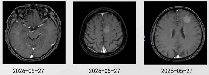
Brain MRI on May 27, 2026, Revealing Newly Developed Brain Metastases
Fourth-Line Treatment: Andaritininib Monotherapy
The patient was hospitalized following disease progression on May 26, 2026. Physical examination showed an ECOG performance status of 1. Brain MRI on May 27, 2026, revealed newly developed brain metastases. The patient had no symptoms related to brain metastases. A biopsy was recommended, but the patient and family again declined.
After fully informing the patient and family and obtaining their informed consent, andaritinib 240 mg once daily (QD) was initiated on May 26, 2026.
Response Assessment: Follow-up chest CT after one month of treatment (June 30, 2026) showed a partial response (PR) in the pulmonary lesions. The PR was maintained at the three-month assessment on August 28, 2026.
Follow-up brain MRI also showed a PR in the brain metastases identified at baseline on May 27, 2026, both after one month of treatment (June 30, 2026) and after three months (August 27, 2026).
As of August 2026, the patient remained on andaritinib treatment and under follow-up. No significant adverse reactions were reported during treatment, and overall tolerability was good.
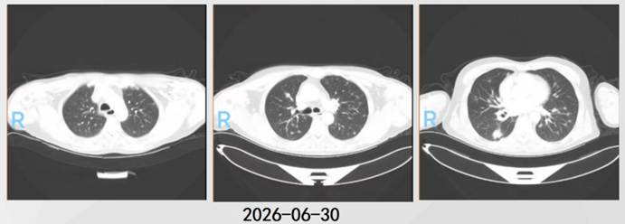
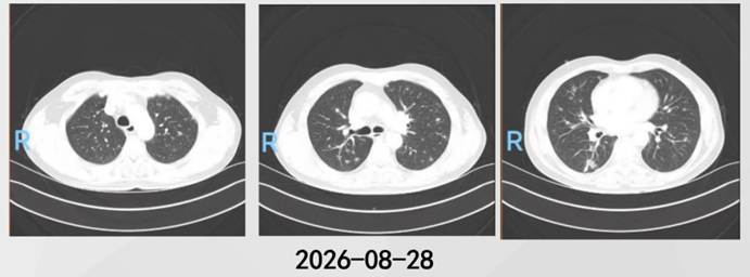
Chest CT on June 30 and August 28, 2026, Showing Tumor Shrinkage
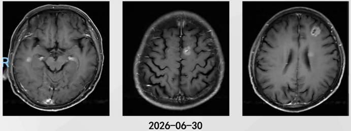
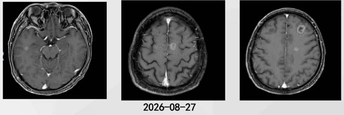
Brain MRI on June 30 and August 27, 2026, Showing Tumor Shrinkage
Expert Commentary
For patients with advanced NSCLC harboring sensitizing EGFR mutations whose disease progresses following first-line treatment with a third-generation EGFR-TKI, clinical decision-making presents multiple challenges. These include substantial heterogeneity in resistance mechanisms, limited access to standard subsequent-line treatment options, and difficulty tolerating intensive treatment regimens in some patients because of prior therapies. In this case, the patient experienced further disease progression after three lines of treatment with aumolertinib, dose-escalated furmonertinib combined with platinum-based chemotherapy, and sacituzumab tirumotecan, followed by the development of new brain metastases. Subsequent-line treatment therefore needed to balance intracranial disease control, antitumor activity, and long-term treatment tolerability.
Andaritininib is a highly selective, irreversible EGFR-TKI. Its medicinal chemistry design incorporates several distinctive features. First, it employs a dual-site binding mode: the 4,6-disubstituted pyrimidine core forms a covalent bond with EGFR C797, while the 1-methylindazole moiety forms hydrogen-bond interactions with residue K745 in the P-loop region. Second, its rigid side-chain design may help improve blood–brain barrier penetration and central nervous system (CNS) exposure. In addition, andaritininib is not a substrate of P-glycoprotein (P-gp) or breast cancer resistance protein (BCRP) and exhibits a low efflux rate, potentially promoting drug retention in brain tissue and increasing CNS exposure [1]. These pharmacological properties warrant attention in patients requiring long-term oral treatment, particularly those whose previous regimens have required repeated adjustments because of tolerability issues.
Andaritininib has received conditional approval from China’s National Medical Products Administration (NMPA) for the treatment of adult patients with locally advanced or metastatic NSCLC harboring EGFR exon 20 insertions (EGFR ex20ins), whose disease has progressed during or after platinum-based chemotherapy, or who are intolerant of platinum-based chemotherapy [2]. The phase II KANNON study [3] has also provided key clinical evidence supporting the use of andaritininib in subsequent-line treatment of advanced EGFR ex20ins-positive NSCLC. The confirmed objective response rate (cORR) was 42.7%, the disease control rate (DCR) was 86.5%, the median duration of response (mDoR) was 8.7 months, and the median progression-free survival (mPFS) was 6.2 months. Among patients with brain metastases at baseline, the cORR assessed by the independent review committee (IRC) was 47.4%, and the DCR was 86.8%, broadly consistent with the efficacy results in the overall population. Andaritininib also demonstrated a favorable safety profile. The incidence of grade ≥3 treatment-related adverse events was 40.2%, with diarrhea (12.0%) and rash (7.6%) being the most common. No cases of interstitial lung disease or grade ≥3 QT interval prolongation were reported in the study [3].
The patient in this case had an EGFR exon 19 deletion (19-Del) mutation and experienced disease progression after multiple lines of treatment. Following a thorough discussion with the patient and family to ensure informed consent, andaritininib was administered, producing encouraging signs of antitumor activity. EGFR 19-Del is a classic sensitizing EGFR mutation, and its kinase domain retains high affinity for ATP-competitive inhibitors. As a highly selective, irreversible EGFR-TKI, andaritininib’s dual-site binding mode may allow it to better accommodate conformational changes in the EGFR kinase domain. In vitro pharmacodynamic data have demonstrated that andaritininib exerts substantial inhibitory activity against 19-Del, L858R, L858R+T790M, and PACC mutations. In nude mouse subcutaneous xenograft models of lung cancer harboring 19-Del or L858R+T790M, andaritininib delayed tumor progression and reduced tumor volume in vivo, demonstrating clear antitumor activity.
Furthermore, the molecular evolution of tumors following EGFR-TKI resistance is highly heterogeneous. T790M mutations, MET amplification, HER2 amplification, and PIK3CA mutations can all serve as mechanisms of acquired resistance [4–5]. Andaritininib also exhibits inhibitory activity against certain resistance-associated mutations, including T790M. This may provide a possible pharmacological explanation for the simultaneous responses observed in the pulmonary and intracranial lesions in this patient during subsequent-line treatment. However, these mechanistic explanations are largely based on previous preclinical studies and the established principles of action of similar agents. The specific inhibitory activity of andaritininib against EGFR 19-Del and secondary resistance mutations still requires validation in larger clinical studies.
Overall, this case describes an individual patient who achieved simultaneous responses in pulmonary and intracranial lesions after receiving andaritininib following multiple lines of treatment, while maintaining good treatment tolerability. Longer follow-up and studies involving larger patient populations are warranted to further clarify differences in targeted therapy sensitivity among EGFR mutation subtypes and to identify the clinical and molecular characteristics of patients who may derive long-term benefit.
Disclaimer:
This article contains information related to prescription medicines and is intended solely for healthcare professionals. This case report describes an individual clinical treatment experience. The patient’s disease characteristics, physical condition, comorbidities, and treatment course were specific to this individual. The treatment outcomes described apply only to this patient and do not imply that the same treatment regimen or medication will produce equivalent efficacy in all patients with similar diseases. Nor does this report constitute diagnostic or treatment advice or a medication recommendation for any specific patient.
The diagnosis and treatment of disease should be determined by qualified healthcare professionals following an individualized assessment of each patient’s circumstances. Readers should not independently modify or adopt any treatment regimen based on this article. This article is intended solely for academic exchange of information regarding recent treatment approaches and does not constitute a recommendation, advertisement, or commercial promotion of any treatment regimen, drug, medical device, or related product. Information about the medical institution and clinical team mentioned herein is provided solely for academic attribution of the case source and does not constitute an endorsement or evaluation of the institution or its medical services. Individuals with health concerns or medical needs should promptly consult a qualified healthcare professional for assessment and care.
This notice is hereby issued.

Professor Lijun Tian