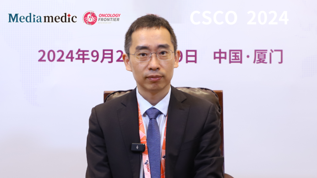
Editor's Note: The 27th Annual Conference of the Chinese Society of Clinical Oncology (CSCO) took place in Xiamen from September 25–29, 2024. This year’s theme, “Patient-Centered, Sharing the Future,” brought together groundbreaking research and developments. Dr. Lei Tang from Peking University Cancer Hospital delivered an insightful presentation on the current state and challenges of imaging in evaluating gastric cancer immunotherapy. In a post-conference interview, he discussed key aspects of imaging evaluation for immunotherapy in gastric cancer.Oncology Frontier: Could you share the main imaging methods and standards currently used in evaluating gastric cancer immunotherapy?
Dr. Lei Tang: With the increasing application of immunotherapy across treatment lines for gastric cancer, imaging has become crucial in assessing therapeutic efficacy, leading to a rise in clinical interest. The widely recognized standard is RECIST 1.1, which categorizes therapeutic responses as CR, PR, SD, or PD based on dimensional measurements. However, in the context of immunotherapy, RECIST 1.1 does not fully meet clinical demands. To address this, modified standards like irRECIST, iRECIST, and imRECIST have been proposed to account for atypical responses seen with immunotherapy, such as pseudo-progression. These adaptations include delayed PD checkpoints (iUPD) and incorporating new lesion measurements for a more objective assessment. Nonetheless, these standards still rely on one-dimensional morphology, which may limit their precision for immunotherapy.
Research in imaging now often explores two main directions: first, new imaging modalities, such as spectral CT, diffusion-weighted MRI, magnetic iron oxide nanoparticle MRI, and PET-based probes, which offer multimodal functional/molecular imaging beyond traditional morphology. The second direction focuses on radiomics and artificial intelligence (AI), linking imaging features to immunologic markers to establish radiomic immune scores (RS/RIS), potentially guiding treatment evaluation and prognosis prediction. Many experts presented their findings at this CSCO conference, showcasing studies on radiomics/AI aimed at enhancing immunotherapy assessment in gastric cancer.
Oncology Frontier: What role does imaging play in monitoring gastric cancer immunotherapy, and what are the future directions?
Dr. Lei Tang: Immunotherapy has improved gastric cancer outcomes, and the need for precise imaging evaluation has grown beyond simple tumor size measurements. For example, as certain locally advanced or even metastatic cases respond well to immunotherapy, surgeons are exploring function-preserving procedures, reducing the scope of gastrectomy, and even considering non-surgical, watch-and-wait approaches. In these scenarios, imaging plays a pivotal role in assessing complete clinical response (cCR) post-immunotherapy. We are exploring new signs to aid in determining cCR after gastric cancer immunotherapy, including markers for identifying immunotherapy-sensitive patients and specific mucosal repair signs for post-treatment cCR. Easily identifiable signs, like “margin signs” or “mucosal line signs,” allow radiologists to interpret images directly without radiomics or AI, facilitating clinical application.
AI in imaging is still developing, and widespread implementation remains challenging due to its limited generalizability across hospitals. In the foreseeable future, while we continue refining AI techniques, traditional imaging features remain valuable. For instance, we’ve developed indicators like the primary lesion area and lymph node short diameter for gastric cancer, combined with intratumoral and intertumoral heterogeneity parameters, to construct models that categorize therapeutic response into three levels. These models have shown a correlation with overall survival (OS) comparable to pathological tumor regression grading (TRG). Calculating primary lesion area improves accuracy by reducing one-dimensional instability, showing strong volume correlation and consistent results. We’ve also integrated body composition analysis, using computer-extracted features like muscle, fat, and peritoneal tissue, to build models that assess immunotherapy efficacy and prognosis, yielding promising results.
In summary, in the era of precision medicine, radiologists’ reports must align with clinical needs, focusing on treatment guidance rather than merely describing observations. Clinical research should adopt a multidisciplinary approach, aiming for multicenter, cross-disciplinary prospective studies to provide high-level evidence to guide practice and improve immunotherapy outcomes for gastric cancer patients.
Dr. Lei Tang
- Chief Physician, Department of Medical Imaging, Peking University Cancer Hospital
- Professor and Doctoral Supervisor
- Executive Member, Gastric Cancer Committee, Chinese Anti-Cancer Association (CACA)
- Leader, Imaging Group, Gastric Cancer Committee
- Key Member, Gastrointestinal Stromal Tumor Committee, Chinese Society of Clinical Oncology (CSCO)
- Published over 30 SCI papers in journals including Annals of Oncology and Radiology


