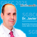
The highly anticipated 19th St. Gallen Breast Cancer Conference (SGBCC) is taking place from March 12 to 15, 2025, in Vienna, Austria, a city renowned for its rich musical heritage. This year’s conference features a special session, "Voice of China," where leading breast cancer experts from CSCO BC share insights into China's clinical, basic, and translational research achievements.Among the distinguished speakers, Dr. Kun Wang from Guangdong Provincial People’s Hospital delivered an outstanding presentation titled: “Radiomics Assists Breast- and Axillary-Conservation After Neoadjuvant Therapy in Breast Cancer .”
Following his presentation, Oncology Frontier had the privilege of interviewing Professor Wang, focusing on how radiomics technology enhances the precision of treatment assessment and outcome prediction in neoadjuvant therapy for breast cancer.
Oncology Frontier: While neoadjuvant therapy (NAT) has increased the rates of breast conservation and axillary preservation, it does not seem to improve overall survival compared to adjuvant therapy. In fact, some studies suggest a higher risk of local recurrence. What could be the underlying reasons for this? Additionally, what are the limitations of relying on pathological complete response (pCR) as the sole criterion for surgical decision-making?
Dr. Kun Wang : One of the most comprehensive systematic reviews on neoadjuvant therapy was the 2018 EBCTCG meta-analysis, published in The Lancet Oncology[1]. This study found no significant difference between neoadjuvant therapy (NAT) and adjuvant therapy (AT) in terms of breast cancer mortality (34.4% vs. 33.7%; HR 1.06, P = 0.31) or overall mortality (40.9% vs. 41.2%; HR 1.0, P = 0.45). However, NAT was associated with a 16% increase in breast conservation rates (65% vs. 49%), which, unfortunately, came with a higher risk of local recurrence (21.4% vs. 15.9%).
Notably, among the clinical trials included in this meta-analysis, two studies enrolled patients who did not undergo surgery after NAT. When these studies were excluded, the issue of local disease control remained an area of concern.
The higher local recurrence rates following NAT can be attributed to multiple factors, including the choice of systemic therapy regimens, challenges in accurately identifying the original tumor bed after treatment, and variability in tumor shrinkage patterns, all of which significantly influence surgical decision-making.
The “No Ink on Tumor” margin guideline, proposed by ASCO/SSO in 2014[2], has become a widely accepted surgical principle. However, applying this concept to post-NAT cases remains a subject of debate due to differences in tumor regression patterns. If a tumor achieves pathological complete response (pCR) following NAT, theoretically, surgery may not be necessary, and biopsy alone could be sufficient for confirmation. For cases with a “fan-shaped” regression pattern, where residual tumor cells are dispersed within the original tumor bed, a wider surgical excision may be required to ensure negative margins. Conversely, the “No Ink on Tumor” principle is more applicable to cases with a “concentric” regression pattern, where the tumor shrinks uniformly towards the center.
Understanding tumor regression patterns is essential for optimizing surgical strategies after NAT. Radiomics holds promise in refining this assessment, aiding clinicians in personalized surgical planning to maximize both oncologic safety and breast conservation outcomes.
Accurately Predicting Tumor Regression Patterns: A Key Clinical Challenge
One of the major challenges in clinical practice is accurately predicting tumor regression patterns following neoadjuvant therapy (NAT). At Guangdong Provincial People’s Hospital, extensive research has been conducted in this field. Currently, the standard assessment tool is MRI, but previous studies, including NRG-BR005 and MICRA trials[3-4], have shown that the negative predictive value (NPV) of imaging-based predictions is only 76%.
By applying radiomics at different stages—before, during, and after NAT—our team has achieved an NPV of nearly 90%. This approach allows for a more precise evaluation of tumor regression patterns, which is crucial for determining surgical resection margins and reducing the risk of positive margins after surgery.
Oncology Frontier: Your team has developed a radiomics-based model that predicts pathological complete response (pCR) while also assessing tumor regression patterns to guide surgical decision-making. Could you share the key indicators of this model and how it performs? What are its unique advantages?
Dr. Kun Wang :Our team began exploring this field in 2017, and in 2019, we published what was likely the first study on radiomics-based pCR prediction in Clinical Cancer Research[5]. This study used baseline MRI data from 586 breast cancer patients to predict treatment response, achieving an area under the receiver operating characteristic (AUC) close to 0.8.
Over time, we recognized that each tumor and each patient responds differently to NAT. This led us to incorporate longitudinal MRI data—analyzing images taken before treatment, during treatment, and toward the end of therapy. By capturing temporal changes in MRI features, we significantly improved the predictive accuracy of our model.
We also integrated machine learning and artificial intelligence to enhance prediction capabilities, which further refined our ability to assess treatment response. This research was later published in eClinicalMedicine[6]. By leveraging these methodologies, we have developed a highly accurate model for predicting pCR and fan-shaped regression patterns, which plays a crucial role in surgical decision-making.
Our current clinical strategy follows a stepwise approach: first, predicting whether pCR can be achieved. Next, identifying whether the tumor exhibits a fan-shaped regression pattern, which would require a wider resection margin. For tumors displaying concentric regression, we apply the “No Ink on Tumor” principle to ensure complete removal while minimizing unnecessary excision.
Oncology Frontier:Radiomics depends on high-quality, standardized imaging data. As this model transitions into clinical practice, how do you address challenges such as variations in MRI equipment parameters across different hospitals and ensuring image segmentation consistency?
Dr. Kun Wang :This is a critical issue. In China, hospitals often use different MRI machines from various manufacturers, each with distinct imaging parameters. To address this, we need to establish standardized protocols to harmonize imaging parameters, ensuring consistency across institutions for reliable model predictions and uniform report interpretation.
Another key aspect is automation. Initially, radiologists needed to manually delineate regions of interest (ROI), extract features, and then make predictions. However, with advancements in artificial intelligence, particularly unsupervised learning, models can now automatically extract features and make predictions in a one-step process. This is the direction we are moving toward, aiming for fully automated, standardized clinical applications.
Oncology Frontier: What is the broader significance of this technology? How do you see its impact on global breast cancer surgery following your presentation at SGBCC?
Dr. Kun Wang :Once this technology becomes fully integrated into clinical practice, it has the potential to eliminate the need for surgery in many patients. If a radiomics-based prediction indicates pCR, patients may only require a core needle biopsy. If the biopsy confirms pCR, there would be no need for further surgery.
For patients with fan-shaped regression patterns, a wider surgical excision might still be necessary to ensure complete removal. However, once this predictive model and treatment strategy gain widespread adoption, breast cancer surgery will become far more precise, leading to better patient outcomes and fewer unnecessary procedures.
Reference:
[1]Early Breast Cancer Trialists’ Collaborative Group (EBCTCG). Long-term outcomes for neoadjuvant versus adjuvant chemotherapy in early breast cancer: meta-analysis of individual patient data from ten randomised trials. Lancet Oncol. 2018;19(1):27-39. doi:10.1016/S1470-2045(17)30777-5
[2]Buchholz TA, Somerfield MR, Griggs JJ, et al. Margins for breast-conserving surgery with whole-breast irradiation in stage I and II invasive breast cancer: American Society of Clinical Oncology endorsement of the Society of Surgical Oncology/American Society for Radiation Oncology consensus guideline. J Clin Oncol. 2014;32(14):1502-1506. doi:10.1200/JCO.2014.55.1572
[3]Basik M,et al. Primary analysis of NRG-BR005, a phase II trial assessing accuracy of tumor bed biopsies in predicting pathologic complete response in patients with clinical/radiological complete response after neoadjuvant chemotherapy to explore the feasibility of breast-conserving treatment without surgery. San Antonio Breast Cancer Symposium 2019. Prog #: GS5-05.
[4]van Loevezijn AA, van der Noordaa MEM, van Werkhoven ED, et al. Minimally Invasive Complete Response Assessment of the Breast After Neoadjuvant Systemic Therapy for Early Breast Cancer (MICRA trial): Interim Analysis of a Multicenter Observational Cohort Study. Ann Surg Oncol. 2021;28(6):3243-3253. doi:10.1245/s10434-020-09273-0
[5]Liu Z., Li Z., Qu J., et al. Radiomics of multiparametric MRI for pretreatment prediction of pathologic complete response to neoadjuvant chemotherapy in breast cancer: a multicenter study. Clin Cancer Res. 2019;25(12):3538–3547. doi: 10.1158/1078-0432.CCR-18-3190.
[6]Huang Y, Zhu T, Zhang X, et al. Longitudinal MRI-based fusion novel model predicts pathological complete response in breast cancer treated with neoadjuvant chemotherapy: a multicenter, retrospective study. EClinicalMedicine. 2023;58:101899. Published 2023 Mar 24. doi:10.1016/j.eclinm.2023.101899


