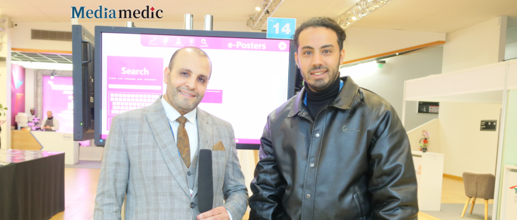
Hepatocellular carcinoma (HCC), the most common primary malignant liver tumor, is one of the leading causes of death in patients with cirrhosis. Non-invasive diagnostic tools such as transient elastography (FibroScan) have shown great promise in identifying patients at high risk of HCC. Recently, the 2025 EASL Liver Cancer Summit was held in Paris, France. Professor Abdel Ghani Atef from the Damas Liver & G.I.T Center in Egypt presented research on the value of ultrasound elastography in the early detection of HCC in patients with cirrhosis. To this end, a journalist from Oncology Frontier traveled to the summit and invited Professor Abdel Ghani Atef to have an in-depth discussion on topics such as the role of FibroScan in HCC screening and the significance of liver stiffness measurement cutoff values.Oncology Frontier: How was the cutoff value of 30.5 kPa determined for early HCC detection, and should this threshold be adjusted for different patient populations such as those with viral hepatitis versus metabolic-associated liver disease?
Professor Abdel Ghani Atef: First of all, we divided our patients into two groups: one group with HCC and one group of cirrhotic patients without HCC. After conducting a statistical analysis and performing FibroScan on both groups, we identified a cutoff value of 30.5 kPa for HCC detection. This threshold was determined based on our statistical findings. Importantly, we did not observe any significant differences in this cutoff among patients with different etiologies, whether related to viral hepatitis or metabolic liver disease.
Oncology Frontier: Your findings indicate that patients with liver stiffness above 25 kPa require close follow-up. What follow-up strategies do you recommend, and should they vary by disease etiology or geography?
Professor Abdel Ghani Atef: During our research, we observed that patients with liver stiffness values between 25 and 30 kPa required close monitoring. For these patients, we recommend a follow-up strategy that includes FibroScan, ultrasound, and alpha-fetoprotein testing every six months. These patients are at a high risk of developing HCC, and our goal is to ensure early detection and treatment. By implementing regular ultrasound and biomarker assessments, we can intervene at an early stage and improve patient outcomes.
Oncology Frontier: How does FibroScan compare to other non-invasive diagnostic tools like FIB-4, GALAD score, and imaging techniques in terms of predictive accuracy for HCC? Could international collaborations help establish a standard multimodal diagnostic model?
Professor Abdel Ghani Atef: There are several non-invasive modalities available for early HCC detection. In our study, we found that FibroScan provides a reliable cutoff of 30.5 kPa for identifying high-risk patients. However, to achieve a more accurate diagnosis, we recommend a multimodal approach that includes alpha-fetoprotein testing, radiological imaging, FibroScan, and the GALAD score. By integrating multiple diagnostic tools, we can improve the precision of HCC detection. Additionally, international collaborations could play a crucial role in validating a standardized multimodal model, ensuring broader applicability and reliability across different populations.
Oncology Frontier: Given the high burden of liver disease in Egypt and China, do you see potential for collaboration in large-scale HCC screening programs? What lessons can both regions share regarding surveillance limitations and early detection strategies?
Professor Abdel Ghani Atef: Yes, collaboration between Egypt and China in this field would be highly valuable. Both countries have a significant number of liver disease cases, and these patients are at increased risk of developing HCC. By working together, we can develop more effective early detection strategies. HCC is a highly aggressive tumor, and early treatment is essential for improving patient outcomes. Establishing large-scale screening programs in both countries would enhance early diagnosis and allow for better treatment planning. Through shared research and surveillance programs, Egypt and China can contribute significantly to the global fight against HCC.
Oncology Frontier: What are the key areas for future research to refine FibroScan’s predictive value for HCC? Do you anticipate advancements in elastography technology to enhance its clinical utility?
Professor Abdel Ghani Atef: Looking ahead, I believe that elastography will continue to be a valuable tool for HCC detection. Since HCC increases liver stiffness, advancements in elastography technology could further improve early detection. In particular, magnetic resonance elastography (MRE) shows great potential due to its high accuracy. However, it remains an expensive option compared to FibroScan. As technology advances, we may see improvements in affordability and accessibility, making MRE a more widely used tool for early HCC diagnosis.